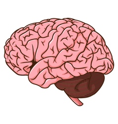
 Everyone knows that the brain has two hemispheres—the right and the left—which perform specific functions. However, the lesser known but equally important organ called the corpus callosum, is essential to communication between the two hemispheres. The corpus callosum is located above the thalamus and under the cortex. As the largest bundle of nerve fibers in the body, it allows the two hemispheres of the brain to connect through neural messages. By providing a highway for communication, the corpus callosum allows the two hemispheres to synchronize and coordinate. For example, the two hemispheres are lateralized; the left hemisphere will control the right side of the body while the right hemisphere will control the left. One interesting demonstration of brain lateralization was an experiment in 1955 by Ronald Meyers, who discovered the function of the corpus callosum. He trained cats to press their nose against the screen when they saw a circle, but not a square. Then, he cut the optical fibers intertwining between the two halves of the brain, but he did not cut the corpus callosum. So, the left eye was connected to the left brain, the right eye to the right, and both brains were connected by the corpus callosum. Then, he trained the cats to do the same experiment, but only with the left eye. Then he tested them, but using the right eye. If the cats completed the experiment successfully, that would mean the information from the left eye had traveled to the left brain, through the corpus callosum and then to the right brain. The cats successfully completed the experiment, proving the corpus callosum connects the two hemispheres. Meyers took his experiment further by using the same experiment, but this time also severing the corpus callosum; those animals failed, since their hemispheres were not able to relay information to each other. Meyers' experiment proves the importance of the corpus callosum; however, what happens if one loses that integral connection between their hemispheres? In the case of some sufferers of epilepsy, surgeons may cut a part of or completely remove the corpus callusum in an effort to stop epileptic discharges from spreading from one side of the brain to the other. Luckily, the brain is a marvelous learning tool and can be taught to function despite obstacles. Those who have their corpus callosum removed, called split-brain patients, relearn how to perform everyday activities. There have been many studies on split-brain patients; one example is the detailed research experiments of Michael Gazzaniga, who has studied split-brain patients for five decades. One such experiment included a patient who looks at a computer screen that is divided in half, so that the right eye could only see the right side of the screen, and the left eye the left screen. Remember, the right eye of each patient is still connected to the left hemisphere, and the left eye to the right. So, when the right screen flashed a picture, the right eye sent the image to the left brain. Since the left brain is verbally dominant, the patient was able to name the picture aloud. However, when the same picture was flashed to the left eye, the left eye sent the image to the right brain, which could not speak the name of the image aloud, but could use the left hand to draw the picture on a piece of paper. Work cited: http://brainmadesimple.com/corpus-callosum.html http://hubel.med.harvard.edu/book/b34.htm http://www.nature.com/news/the-split-brain-a-tale-of-two-halves-1.10213 https://m.youtube.com/watch?feature=youtu.be&v=zx53Zj7EKQE 
0 Comments
Leave a Reply. |
AuthorHello. Welcome to all things psychology. Enjoy! Archives
December 2017
Categories
All
|
 RSS Feed
